| Patient D., 56. Longitudinal section: stenosis of the abdominal aorta as a result of a blood clot, which is located next to the partially calcined atheromatous plaque. | |
 |
 |
| Patient S., 48 years old. Cross section: swelling of the gallbladder wall in normal liver parenchyma. The patient has acute hepatitis. | |
 |
 |
| Patient B., 44 years old. Micronodular cirrhosis. | |
 |
 |
| Patient Yu 8 years. Longitudinal section: Ascaris in the common bile duct. | |
 |
 |
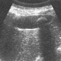
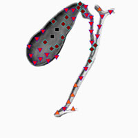
| Patient P., 36 years. The gall bladder contains a large single calculus. | |
 |
 |
| Patient S., 84 years old. Atrophy of the pancreas tail. | |
 |
 |
| Patient A., 68 years old. Longitudinal sections: cystadenocarcinoma pancreas. | |
 |
 |
| Patient G., 34 years old. Cross section: erosive gastritis. | |
 |
 |
| Patient E., 42 years old. Longitudinal section: multiple kidney cysts | |
 |
 |
| Patient G., 34 years old. Cross section: erosive gastritis. Patient B., 68 years old. Cross section: swelling of the lower pole of the left kidney, germinating in the left ureter is the cause of obstructive hydronephrosis. | |
 |
 |
| Patient S., 56 years old. Longitudinal section: the right kidney stone | |
 |
 |
| Patient I., 65 years old. Longitudinal section: a benign adenoma in the right adrenal gland. | |
 |
 |
|
Patient K., 38 years old. Chronic bladder inflammation (chronic cystitis). |
|
 |
 |
| Patient G., 34 years old. Cross section: varicocele with multiple dilated veins. | |
 |
 |
| Patient T., 46 years old. Longitudinal section: a large tumor (cancer) of the cervix. | |
 |
 |
| Patient M., 32 years old. Cross section: endometrioma. | |
 |
 |
| Patient D, 41 years old. Cross section: ovarian cyst with irregular contours. | |
 |
 |
| Patient N., 45 years old. Paramedian herniation of intervertebral disc with the right-lateralization. | |
 |
 |
| Patient S., 68 years old. Osteoblastic metastatic carcinoma of the prostate gland in the vertebrae. | |
 |
 |
| Patient K., 74 years old. Metastasis of kidney cancer in the lumbar vertebrae. | |
 |
 |
|
Patient N., 24 years old. Compression body fracture of the first lumbar vertebra .. |
|
 |
 |
| Patient D., 57 years old. Neurofibromatosis. | |
 |
 |
| Patient B., 37 years old. Lipoma of the hypothalamus. | |
 |
 |
| Patient C., 54 years old. Encephalopathy (chronic cerebrovascular insufficiency). | |
 |
 |
| Patient N., 43 years old. Acute ischemic stroke. | |
 |
 |
| Patient T., 38 years old. Subfrontalny meningioma. | |
 |
 |
| Patient Z., 62 years old. Metastasis of renal adenocarcinoma in the right hemisphere of the cerebellum. | |
 |
 |
| Patient Z., 62 years old. Metastasis of renal adenocarcinoma in the right hemisphere of the cerebellum. | |
 |
 |
| Patient P., 47 years. Typical supratentorial lesions in multiple sclerosis | |
 |
 |
| Patient E., 54 years old. Osteochondrosis of the lumbar spine. | |
 |
 |
| Patient B., 56 years old. Osteochondrosis of the cervical spine with a reduction in height of the disc and rear hernias. | |
 |
 |
| Patient J., 67 years old. Fibrosarcoma sacrum. | |
 |
 |
| Patient A., 58 years old. Simpatoblastoma cervical localization. | |
 |
 |
| Patient A., 58 years old. Simpatoblastoma cervical localization | |
 |
 |
| Patient I., 49 years old. Perineural arachnoid cysts. | |
 |
 |


